Wobbler syndrome in horses, known to vets as cervical vertebral malformation (CVM), is a neurological disease in which the bones of the neck compress the spinal cord and rob the horse of coordination. The result is the unsteady, swaying, slightly drunk-looking gait that gives the condition its name, and it almost always shows up in the hind legs first. It is the one spinal cord disease in the horse for which surgery is performed routinely, and it is also one of the most commonly misread, because the earliest signs look far more like clumsiness or bad schooling than like a neurological emergency.
This guide covers what the condition actually is, the two forms it takes, how quickly it progresses, what your vet is looking for during a neurological workup, and what the realistic outcomes are from both conservative management and surgery. Where a figure comes from a specific study, it is named, because a great deal of what circulates about wobblers online is confidently quoted and poorly sourced.
Table of Contents
What Is Wobbler Syndrome in Horses?
Wobbler syndrome describes the clinical picture produced by compression of the spinal cord inside the neck. The cervical vertebrae form a bony tunnel through which the spinal cord runs. If that tunnel is too narrow, or if two vertebrae move against each other more than they should, the cord gets squeezed. Nerve signals travelling between the brain and the limbs are degraded, and the horse loses the fine positional awareness it needs to place its feet accurately.
That loss of positional sense is called ataxia, and it is the single defining feature of the disease. A wobbler is not weak in the sense of being tired or unfit, and it is not lame in the orthopaedic sense. It genuinely does not know precisely where its feet are. That is why a wobbler will scuff a hind toe on flat ground, cross its legs on a tight turn, or stand with its feet in an odd position and leave them there.
Hind limbs are affected first and more severely in the great majority of cases, because of how the nerve tracts serving the hind limbs are arranged within the cord. Front limb signs appear later and usually indicate a more advanced or higher lesion.
The Names Vets Use: CVM, CVCM, CVSM and CSS
The naming is genuinely inconsistent across the literature, and it causes real confusion for owners reading around a diagnosis. All of the following refer to the same broad disease:
- CVM, cervical vertebral malformation. The commonest umbrella term.
- CVCM, cervical vertebral compressive myelopathy. Emphasises that the cord is being compressed. Widely used in current veterinary literature.
- CVSM, cervical vertebral stenotic myelopathy. Very common in North American sources and in the surgical literature.
- CSS, cervical static stenosis. Refers specifically to the static form, not to the disease as a whole.
If your vet uses one term and the referral report uses another, that is not a disagreement about the diagnosis. It is just an unsettled vocabulary. Our glossary entry on equine wobbler syndrome covers the terminology in more detail.
Equine Wobbler Syndrome Is Not the Same Disease as Canine Wobbler Syndrome
Worth clearing up early, because a large share of the search traffic for the bare phrase “wobbler syndrome” is about dogs. Dobermanns and Great Danes get a condition also nicknamed wobbler syndrome, properly cervical spondylomyelopathy. The mechanism rhymes, in that the neck compresses the cord, but the breeds, the age of onset, the imaging and the surgical options are all different. Do not apply canine advice, drug doses or prognosis figures to a horse.
Type 1 vs Type 2: The Two Forms of Cervical Vertebral Malformation
The distinction between the two forms matters more than any other single piece of information in a wobbler workup, because it changes the treatment plan, the surgical options and the prognosis.
Type 1: Cervical Vertebral Instability (the Dynamic Form)
In the dynamic form the spinal canal is broadly wide enough at rest, but there is excessive movement between two adjacent vertebrae. When the horse flexes its neck, the vertebrae shift out of alignment and pinch the cord. Release the flexion and the compression eases.
- Typically affects young, rapidly growing horses, most often between about six months and three years.
- Lesions cluster in the mid neck, commonly C3 to C4 and C4 to C5.
- Over-represented in Thoroughbreds and tall continental warmbloods.
- Signs often fluctuate, appearing worse after exercise or on a day the horse has been working on a tight circle.
Because compression is intermittent, this is the form most likely to respond to management that buys time while the skeleton matures, and the form most suited to surgical fusion, which abolishes the abnormal movement.
Type 2: Cervical Static Stenosis
In the static form the canal is permanently too narrow, so the cord is compressed whether the neck is flexed, extended or neutral. The narrowing comes from malformed vertebral bone, from thickened joint capsules, or from arthritic enlargement of the articular process joints.
- Affects older horses, frequently from around eight years upwards, though it is also seen in young horses with severe malformation.
- Lesions sit lower down the neck, commonly C5 to C6 and C6 to C7.
- Often accompanies degenerative joint disease of the neck joints.
- Signs are usually slowly and steadily progressive rather than fluctuating.
Plenty of horses show features of both forms at once, and multiple compression sites are common. That is one reason imaging interpretation belongs with a clinician who does neurological work regularly.
What Age Does Wobbler Syndrome Start?
There is no single answer, because the two forms have different age profiles, and that is exactly why the question generates so much conflicting advice.
- Six months to three years is the classic window for the dynamic form. Many cases are picked up as the horse is broken or enters pre-training, simply because that is the first time anyone asks it to work in a controlled way and watches it move.
- Eight years and older is typical for the static, arthritic form.
- Trauma can produce a wobbler-like picture at any age. A horse that has flipped over backwards or come down hard in the field can compress its cord without any developmental malformation at all.
In one surgical case series the median age at surgery was 24 months, with a range from 12 months to 168 months, which is a fair illustration of how wide the real spread is (Pezzanite et al., Equine Veterinary Journal, 2022).
Which Horses Are Most at Risk?
Breed and Sex
Wobbler syndrome is over-represented in Thoroughbreds, Tennessee Walking Horses, Quarter Horses and tall warmblood types such as the Holsteiner. In UK racing yards most cases surface between weaning and the start of pre-training.
The sex bias is strong and well documented. Reported prevalence in male horses runs at roughly 75 to 80 per cent of cases, and prevalence in Thoroughbreds as a breed has been reported at 1.3 to 2 per cent (Pezzanite et al., 2022). Colts grow faster and reach greater body weight at the same age, which is the most widely accepted explanation, though it is not the only one proposed.
Growth Rate and Nutrition
Wobbler syndrome sits within the broader group of developmental orthopaedic diseases, alongside osteochondrosis dissecans (OCD), physitis and angular limb deformities. The shared risk factors are rapid growth, high-energy feeding and imbalanced trace minerals, in particular copper and zinc and a distorted calcium to phosphorus ratio.
The practical point for anyone breeding or raising youngstock is that fast growth is not the same as good growth. A yearling carrying too much condition on a high-starch ration is at greater risk than one grown steadily on forage and a properly balanced mineral supply. Our guides to equine diet and nutrition and weaning a foal successfully cover the feeding side in more depth.
If a foal or yearling already shows any developmental orthopaedic disease, it is reasonable to ask your vet to assess the neck as well as the limbs.
Is Wobbler Syndrome in Horses Genetic?
A heritable component is strongly suspected but no single causative gene complex has been mapped, and wobblers is not a condition you can currently screen for with a DNA test. What the evidence supports is that susceptibility is inherited as a tendency, and that the tendency then interacts with growth rate and nutrition to produce disease or not.
The sensible breeding position is therefore cautious rather than absolute. If a mare has produced an affected foal, repeating the same mare and stallion combination is a real gamble, and it is worth knowing whether a stallion has affected offspring on the ground before committing.
Early Signs of Wobbler Syndrome in Horses
Early cases are routinely dismissed as clumsiness, laziness behind, or a schooling problem. Catching it early genuinely changes the options available, so these are the things worth taking seriously:
- Toe dragging behind. Look at the hind shoes. Squared-off, scuffed toes within a fortnight of shoeing is a hard physical sign, and your farrier will often notice it before you do.
- Crossing over or pivoting. Asked to turn tightly, the horse swings a hind leg across the midline, or plants a hind foot and pivots on it instead of picking it up and placing it.
- An odd stance. Standing base-wide for stability, or leaving a foot in a strange position after moving and not correcting it.
- Knuckling. The fetlock buckles forward briefly as the horse moves off, particularly after standing.
- Trouble reversing. Reluctance to rein back, dragging the hind feet backwards, or stumbling in reverse.
- Disproportionate difficulty on slopes and ramps. Loading and unloading can become a sudden problem.
- Bunny-hopping behind at canter, or persistent trouble holding a canter lead.
- Apparent lameness that does not block out. A gait abnormality that fails to respond to diagnostic nerve blocks is a strong hint that the problem is neurological rather than orthopaedic. Our guide to lameness in horses explains how that distinction is drawn.
One caution worth stating plainly: none of these signs is unique to wobblers. Each of them has orthopaedic and neurological differentials, which is why the workup matters more than the sign list.
What a Wobbler Actually Looks Like
A very large share of the searching around this condition is people looking for video and photographs, and the reason is obvious: ataxia is a movement pattern, and movement patterns are hard to convey in prose. If you are trying to work out whether what you are seeing in your own horse counts, a few points help.
- Ataxia is most visible at the walk, not the trot. Trot can mask mild ataxia because the increased impulsion carries the horse through the placement error.
- It becomes obvious under manoeuvres that remove the horse’s ability to compensate: tight circles, walking on a slope, backing up, walking with the head elevated, or walking over a low kerb.
- It is consistent but variable in degree. A horse does not have ataxia on one circle and not the next, but it may look markedly worse on a slope than on the flat.
- Film it. A phone video of the horse walking away from and towards the camera on a hard, level surface, then in both directions on a tight circle, then backing up, is genuinely useful to a vet and costs nothing. Take it before the visit.
Do not attempt tail-pull or blindfold tests yourself. They are part of a veterinary examination for good reason, and an ataxic horse pulled off balance can fall on the person doing the pulling.
How Fast Does Wobbler Syndrome Progress?
This is one of the most common questions and one of the least well answered elsewhere. The honest answer is that progression is variable and poorly predictable, but it does follow recognisable patterns.
- The dynamic form in a young horse often progresses over weeks to a few months, and may then plateau. Some cases stabilise as growth slows and skeletal maturity arrives, which is the entire rationale behind conservative management.
- The static, arthritic form in an older horse typically creeps, worsening over months to years as the arthritic change advances.
- Traumatic cases are sudden by definition, appearing over hours to days.
- Sudden, sharp deterioration in a previously stable horse is a reason to call the vet promptly rather than watching it. It may reflect an acute injury on top of existing narrowing, or it may point away from wobblers entirely towards an infectious cause.
What progression does not usually do is reverse on its own. A horse that appears to improve spontaneously is more likely to be having a good day, to be moving on better footing, or to have been misdiagnosed in the first place. That is why grading at repeated intervals by the same vet, ideally with video, is more informative than any single assessment.
The Mayhew Ataxia Grading Scale
Vets grade ataxia on the Mayhew scale, running from 0 to 5. Knowing it lets you have a much more precise conversation about whether your horse is stable, improving or deteriorating.
- Grade 0. Normal. No deficits detectable.
- Grade 1. Deficits just detectable at the walk, and only revealed by manoeuvres such as tight circles, backing up, or walking on a slope.
- Grade 2. Deficits easily detected at the walk, and clearly exaggerated by manoeuvres.
- Grade 3. Deficits prominent at the walk without any manoeuvre. The horse may stumble or buckle, but generally stays upright.
- Grade 4. Stumbling and falling at the walk. The horse is a clear danger to itself and to anyone handling it.
- Grade 5. Recumbent and unable to rise. A welfare emergency.
Grade is one input to the surgical decision rather than a fixed cut-off. The surgical series described further down operated on horses graded 1 to 3 (Pezzanite et al., 2022), and the horse’s age and the number of compression sites also weigh in (UC Davis Center for Equine Health). Reported improvement is usually one or two grades, so a horse at grade 4 or 5 is unlikely to regain normal neurological function (UC Davis). Barrie Grant, who helped develop the operation, writes that a horse above grade 3 with obvious compression low in the neck has a very poor chance of responding to medication, time, diet and rest, and that euthanasia is indicated for a grade 4 or 5 horse whose owners do not want to attempt surgery (Barrie Grant on medical treatment).
The scale is also the standard yardstick for reporting outcomes, so when a study says a horse “improved by one grade”, this is the scale it means. Our companion article on ataxia in horses covers grading and the wider differential list in more depth.
How Wobbler Syndrome Is Diagnosed
Diagnosis is a sequence, not a single test, and no one step is sufficient on its own. That point is worth holding onto, because the most common diagnostic error in wobblers is treating a neck radiograph as a verdict.
Neurological Examination
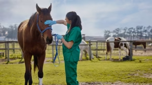
The vet watches the horse move on level ground, on a tight circle, on a slope and over a kerb, then tests responses that reveal deficits the horse can otherwise mask. Typical components include the tail pull at rest and at walk, walking with the head elevated, backing up, and circling in both directions. The aim is to establish first that the horse is genuinely ataxic rather than lame, and second where in the nervous system the lesion is likely to sit.
A neurological examination also grades severity, which sets the baseline against which everything afterwards is measured.
Radiographs and the Sagittal Ratio
Lateral radiographs of the neck are the usual first imaging step and can be taken with the horse standing. Beyond looking for obvious malformation and arthritic change, radiographs allow calculation of the sagittal ratio: the minimum height of the spinal canal divided by the width of the vertebral body at the same site. Dividing by the vertebral body corrects for the size of the horse and for magnification, so the ratio is comparable between animals.
The cut-off values quoted vary between studies, and it is worth knowing that rather than treating one number as definitive:
- The original description by Moore and colleagues (1994) is the source of the widely quoted thresholds of roughly 0.52 in the mid cervical vertebrae and 0.56 at C7.
- A later assessment by Hahn and colleagues (2008), published in Veterinary Radiology and Ultrasound, found that horses with a ratio of 0.485 or below at any site could be correctly classified as affected, and that the site with the smallest intervertebral ratio was always the site where the cord was actually compressed. The same study found that intravertebral measurements alone could not reliably predict where the compression was.
The practical conclusion from that work is the important part: sagittal ratios narrow the field, they do not close it, and they are most accurate when combined with myelographic measurements rather than used alone. A horse can have a suggestive ratio and no meaningful compression, or a borderline ratio and a significant lesion.
Myelography, CT and MRI
Myelography remains the traditional confirmatory test. Contrast medium is injected into the space around the spinal cord under general anaesthesia, and radiographs are taken with the neck neutral, flexed and extended. A reduction of more than about 50 per cent in the dorsal contrast column at a given site is the conventional threshold for significant compression, and comparing flexed with neutral views is what separates dynamic from static lesions.
Computed tomography, increasingly with contrast, has become the preferred approach at centres that have the equipment, because it resolves the lower neck far better than plain films and removes much of the guesswork about which site is the real one. In the UK, Rainbow Equine Hospital lists CT myelography among its wobbler workup services.
Both approaches require general anaesthesia, which in an ataxic horse carries a recognised risk during recovery. That risk is part of the decision, not a footnote to it.
Cerebrospinal Fluid Analysis
Cerebrospinal fluid is usually unremarkable in a straightforward wobbler. It is sampled not to confirm CVM but to exclude the infectious and inflammatory causes that can imitate it, which is a different and equally important job.
What Else Could It Be? Differentials to Rule Out
Spinal cord compression is only one of several conditions that produce an ataxic horse, and some of the alternatives are contagious, treatable, or both. A vet will work through a list along these lines:
- Equine herpesvirus myeloencephalopathy (EHM), the neurological form of equine herpesvirus. Often febrile, sudden in onset, and a biosecurity emergency for the whole yard. See also our overview of equine herpesvirus.
- Equine protozoal myeloencephalitis (EPM), caused by Sarcocystis neurona. Common in the Americas, rare in the UK, and worth raising if the horse has been imported.
- Equine degenerative myeloencephalopathy and vitamin E deficiency, which is linked to vitamin E status and typically presents in young horses.
- Cerebellar abiotrophy, which produces cerebellar signs rather than the placement deficits of a wobbler.
- Botulism, which causes weakness and tremor rather than true ataxia.
- Tetanus, which causes generalised stiffness and spasm and is unmistakable once established.
- Cervical arthritis without cord compression, which is a common incidental finding in older horses and needs imaging to distinguish.
- Spinal trauma from a fall, a somersault or going over backwards.
- West Nile virus, which is notifiable in the UK and a genuine differential in the right geography.
In South Africa, African horse sickness belongs on that list as well.
The trigger to prioritise infectious causes ahead of anaesthesia is straightforward: a fever, a recent move, or any known outbreak on the yard. Otherwise, in a routine workup the cerebrospinal fluid sample and the blood and fluid tests for infectious causes are taken during the myelogram’s anaesthesia (Barrie Grant’s diagnostic sequence).
Treatment Options for Wobbler Syndrome in Horses
Treatment splits into three approaches, and which is appropriate depends on the form, the grade, the age of the horse and the number of compression sites.
Conservative Management and the Paced Diet
For young horses caught early, the established conservative approach is a restricted, paced diet combined with confinement. The logic is to slow growth deliberately so that the cervical anatomy can mature without the cord being compressed further during the fastest growth phase.
The protocol traces to Donawick, Mayhew, Galligan and colleagues (1989), working at the New Bolton Center of the University of Pennsylvania. In its original description it applies to foals under a year old, and preferably diagnosed before six months. In outline it involves weaning if the foal is still suckling, removing creep feed, restricting energy and protein while maintaining or increasing trace minerals through a low-energy ration balancer, and confining the foal with only limited controlled turnout.
Two honest caveats, both of which are routinely omitted elsewhere. First, there are no controlled studies establishing the efficacy of the paced diet or of anti-inflammatory protocols in this disease, so the widely circulated success percentages should be treated with scepticism. Second, the cost is real: affected foals look poor as yearlings, and breaking and training are set back by months. That is a defensible trade for a horse that recovers, and a significant loss for one that does not.
Anti-Inflammatory and Joint Medication
Non-steroidal anti-inflammatories reduce cord and soft tissue inflammation and often produce a modest, temporary improvement. Corticosteroid injection of the cervical articular process joints, usually placed under ultrasound guidance, is used particularly in the older arthritic cases, and can improve neck comfort.
Set expectations carefully here. Medication addresses inflammation, not the underlying bony narrowing or instability. Improvement in neck comfort is not the same as resolution of ataxia, and a horse that becomes more comfortable but remains ataxic is no safer to ride than it was before.
Surgery: Cervical Vertebral Interbody Fusion
The established surgical treatment is cervical vertebral interbody fusion. A hollow, perforated, threaded implant, the Bagby basket, is packed with bone graft and screwed into an osteotomy between two adjacent vertebrae. Bone grows through the perforations, the two vertebrae fuse into one rigid unit, and the abnormal movement that was pinching the cord is abolished.
The implant was invented by Dr George Bagby and developed for equine use with Dr Barrie Grant and colleagues at Washington State University in the late 1970s (Washington State University). It became widely known outside the profession when the technique was used on the champion Thoroughbred Seattle Slew in 2000, which is why the implant is still often called the Seattle Slew basket (Barrie Grant’s account of the procedure).
One point of terminology worth getting right, because it is frequently muddled: interbody fusion is not laminoplasty. Fusion deliberately eliminates movement between two vertebrae. Laminoplasty reshapes bone to enlarge the canal while preserving motion. They are different operations with different indications, and a horse is not a candidate for both interchangeably.
Surgery is not widely available. In the UK it is performed at a small number of centres, and Rainbow Equine Hospital describes its team as one of the only centres in the country offering it.
Newer Surgical Constructs
Newer approaches add a polyaxial pedicle screw and rod construct, borrowing hardware concepts from human spinal surgery, to improve stability at the fusion site. A case series of ten horses operated between 2015 and 2019 gives a realistic picture of what modern surgery achieves and costs the patient (Pezzanite et al., 2022):
- Median age 24 months. Pre-operative ataxia grade 1 to 3. Fusion at one site in three horses and two sites in seven.
- Two of the ten horses were euthanised within the first year.
- Of the eight with at least a year of follow-up, six improved by one to three grades, with an average improvement of 1.25 grades.
- Four horses improved to grade 0 or 1. In two, the gait was unchanged but neck comfort improved.
- Complications were common though mostly manageable: seroma formation in nine of ten, pain in five, fever in four, and screw breakage in two. No fatal complications related to implant placement occurred.
The authors’ own conclusion is measured, and it is the right note for owners as well: the technique produced at least one grade of improvement in six of ten horses operated, which is similar to other methods rather than a breakthrough. Surgery is a genuine option that helps many horses. It is not a cure, and it is not a certainty.
Long-Term Prognosis and Return to Work
Prognosis turns on how severe the signs are, how long they have been present and the level of work expected afterwards, and younger horses with mild signs of short duration do best (UC Davis Center for Equine Health). Duration matters for surgery too: in an Ohio State series of 73 horses, those that regained athletic function or improved by two or more grades had shown signs for a shorter time before the operation, while the number of compression sites did not affect the long-term result of fusion (Moore et al., 1993). Static lesions low in the neck are the hardest to treat surgically (Barrie Grant on prognosis). A two-year-old at grade 1 whose signs began a few weeks ago is a different proposition from a ten-year-old at grade 3 with long-standing static stenosis low in the neck.
The blunt statistic worth knowing is that two US hospital studies found between 38 and 65 per cent of horses with CVM were euthanised: 172 of 263 horses in a multicentre study (Levine et al., 2010), and 33 of 87 Thoroughbreds with a presumptive diagnosis, who were euthanised rather than discharged for conservative treatment (Hoffman and Clark, 2013). The range is often quoted as 33 to 66 per cent (cited in Pezzanite et al., 2022), but 33 is the number of horses the second study euthanised, not a percentage. That is a wide range, reflecting differing case mixes and differing thresholds for intervention, but the message is consistent: this is a serious diagnosis with a genuine mortality attached.
For horses that do improve, improvement is usually measured in grades rather than in a return to normal. A horse that goes from grade 3 to grade 1 is a substantially better and safer animal, but it has not become neurologically normal. Recovery after fusion is measured in many months, and no ridden work should be contemplated until the surgeon confirms the fusion is solid and the horse has been re-graded.
Retired wobblers can make good companions, and horses that improve to grade 0 or 1 sometimes return to work. Both outcomes are legitimate. Neither should be assumed at the outset.
What Wobbler Treatment Costs, and What Insurance Covers
Costs are genuinely difficult to state, and anyone quoting you a confident single figure for the UK or South Africa is guessing. Referral hospitals do not publish wobbler surgery pricing, it varies with the number of sites fused, the imaging required and the length of hospitalisation, and it changes year to year.
What is reliable is the shape of the spend, so budget across all of it rather than for the operation alone:
- Initial workup: neurological examination and standing radiographs.
- Referral imaging: CT myelography or myelography, including general anaesthesia.
- Surgery and hospitalisation, if you proceed.
- Follow-up imaging to confirm fusion.
- Twelve months or more of rehabilitation and keep, during which the horse earns nothing.
Ask for a written estimate covering every stage before committing to the first one, and ask specifically what happens to the money already spent if imaging shows the horse is not a surgical candidate.
On insurance, treat this as a serious practical constraint rather than an afterthought. Developmental and congenital conditions are commonly excluded from equine policies, and once wobbler syndrome is on a horse’s record it becomes very difficult to insure for loss of use. Read your own policy wording before the workup rather than after it. If you are buying, a neurological assessment belongs in the pre-purchase vetting checklist, particularly for a young horse of a predisposed breed.
Can Wobbler Syndrome in Horses Be Prevented?
You cannot eliminate a condition with an inherited component, but you can avoid loading the dice against a young horse:
- Feed broodmares a properly balanced ration through late gestation, with attention to trace minerals rather than just calories.
- Wean onto a forage-first diet rather than high-starch creep feed, and aim for steady growth instead of maximum growth.
- Monitor growth and condition. A yearling carrying excess condition is worth a conversation with your vet or a nutritionist. Our guide to feeding horses for weight gain covers the same balance from the other direction.
- Think carefully before repeating a mare and stallion cross that has already produced an affected foal.
- Have any young horse with persistent clumsiness, or with other developmental orthopaedic signs, assessed neurologically before it goes into work rather than after a rider has fallen off it.
General welfare and health management guidance for owners is available from the British Horse Society, and BEVA maintains guidance for the veterinary profession.
Welfare, Safety and the Decision to Retire
Wobbler syndrome is a rider safety issue as much as a clinical one, and that deserves stating without euphemism. A horse that cannot reliably place its feet may fall, and it may fall with someone on it or beside it. Ataxic horses do not stumble predictably, and they cannot always catch themselves.
In practice, grade 1 horses are sometimes ridden on flat, even ground after an explicit conversation with the vet about risk. From grade 2 upwards, ridden work is difficult to justify. Grade 4 and 5 cases are welfare emergencies.
Owners in the UK have a legal duty of care under the Animal Welfare Act 2006, and in South Africa under the Animals Protection Act 71 of 1962. Both frame the same question: whether continuing to use the horse is compatible with its welfare. Retirement to companion status is often the right answer, and for severely affected horses euthanasia is the humane one.
If you suspect wobbler syndrome, get a vet out rather than waiting to see whether the horse grows out of it. Early assessment materially changes what is possible, and a firm diagnosis, even an unwelcome one, is better than months of guessing.
Frequently Asked Questions
Can a horse with wobbler syndrome recover?
Recover is the wrong yardstick. Horses improve rather than recover, and improvement is measured in ataxia grades. In a recent surgical series, six of eight horses followed for at least a year improved by one to three grades, and four reached grade 0 or 1. Some young horses managed conservatively stabilise as they mature. What effectively never happens is a return to guaranteed neurological normality, because the underlying bony change does not reverse.
What is the life expectancy of a horse with wobbler syndrome?
It depends almost entirely on severity. Mild, stable cases can live a normal lifespan as companions or, in some cases, in light work. Severe cases have a poor outlook, and two US hospital studies found that between 38 and 65 per cent of horses with the disease were euthanised. Grade at diagnosis, and whether the horse is deteriorating, matter far more than age.
Can you ride a horse with wobbler syndrome?
Only after an explicit risk conversation with your vet, and generally only at grade 1 on flat, even going. From grade 2 upwards the rider is in real danger, because an ataxic horse can fall without warning and cannot reliably recover its balance. A horse that has been surgically treated and re-graded to 0 or 1 may return to work, but only once the surgeon confirms fusion is solid.
How is wobbler syndrome in horses treated?
Three routes. Young horses caught early may be managed with a restricted, paced diet and confinement to slow growth. Anti-inflammatory medication and corticosteroid injection of the neck joints can improve comfort but do not address the compression. Cervical vertebral interbody fusion, using a Bagby or Seattle Slew basket, surgically abolishes the abnormal movement at the affected site and is the only treatment that addresses the mechanical cause directly.
Is wobbler syndrome the same as EPM?
No. Wobbler syndrome is mechanical compression of the spinal cord by the bones of the neck. Equine protozoal myeloencephalitis is an infectious disease caused by the parasite Sarcocystis neurona, common in the Americas and rare in the UK. They can look similar on a first examination, which is exactly why imaging and cerebrospinal fluid analysis are used to separate them.