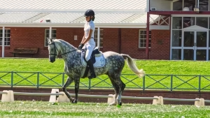
Melanoma in horses – specifically the small, black, dome-shaped tumours of the skin and pigment-producing cells – is one of the most common cancers in equine medicine. The statistics are striking: peer-reviewed research consistently finds that more than 80% of grey horses aged 15 years and older develop at least one melanoma in their lifetime. For most of those horses the tumours are slow-growing nuisances rather than killers, but a significant minority become locally invasive, ulcerated and ultimately malignant. This guide explains the underlying graying genetics (the Syntaxin 17 gene), the four clinical patterns described in the Pimenta 2023 narrative review, how UK and SA veterinary surgeons grade and treat melanoma in 2026, and how to decide whether to buy a grey horse with existing melanomas.
Table of Contents
What Is Equine Melanoma?
Equine melanoma is a tumour that arises from melanocytes – the cells in the skin that produce the pigment melanin. The tumours typically appear as firm, black, smooth-surfaced nodules in non-haired areas of skin, most often under the tail, around the anus and external genitalia, on the lips, eyelids and parotid (jowl) region. A nodule sitting on the eyelid margin rubs the ocular surface with every blink, so a grey horse with a weeping eye deserves a proper look rather than being written off as showing the usual signs of pink eye in horses.
The condition has been recognised in horses for more than two centuries. What has changed in the last decade is our understanding of why grey horses are at extreme risk – and what we can do about it once tumours appear. The two pillars of modern equine melanoma medicine are early intervention rather than “watch and wait”, and the use of the canine Oncept vaccine off-label in horses.
Why Grey Horses? The Syntaxin 17 Story
The connection between grey coat colour and melanoma was confirmed at the genetic level in 2008 by Pielberg et al., who identified a 4.6 kb duplication in intron 6 of the Syntaxin 17 (STX17) gene on equine chromosome 25 as the cause of progressive depigmentation in grey horses. Every grey horse on the planet today carries this mutation; it is dominant, which is why a grey horse always produces at least one grey foal when bred to a non-grey.
The same mutation that causes greying also drives melanocyte proliferation. As the coat fades from coloured to grey to white, melanocytes elsewhere in the body – particularly in the skin around the anus and genitals – begin to over-proliferate, forming the characteristic dark nodules. The horse’s pigment cells are not destroyed during greying; they are simply prevented from making their way to the hair shaft. Those cells accumulate, and a proportion of them turn neoplastic.
More recent work (Brodesser et al. 2025; Pimenta et al. 2023) has identified additional modifier loci including ASIP and NR4A3 that influence which grey horses develop melanoma earliest and which progress to malignancy. The genetic picture is now well enough understood that future genetic testing for melanoma risk is plausible – though not yet commercially available.
The Four Clinical Patterns
Modern equine oncology classifies melanocytic tumours into four clinical patterns (Pimenta et al. 2023). Knowing which pattern your horse has informs the prognosis and treatment plan.
1. Melanocytic Naevus
The classic benign “mole”. Small, well-circumscribed, slow-growing. Usually appears in younger horses (under 6 years). May appear on any colour of horse. Generally non-aggressive; surgical removal is curative.
2. Discrete Dermal Melanoma (Grey Horse Melanoma)
The form seen in adult grey horses. Multiple, well-encapsulated, dermal nodules at the classic sites – under the tail, perineum, sheath, eyelids. Most cases are slow-growing for years before either stabilising or transitioning to a more aggressive form. The 80% lifetime risk in grey horses 15+ refers principally to this pattern.
3. Dermal Melanomatosis
An aggressive variant in which the dermal melanomas become confluent, ulcerated, and grow into large coalescing masses. The lesions interfere with defecation, sweating, mating or harness fitting. This pattern often requires emergency surgical debulking and carries a guarded prognosis.
4. Anaplastic Malignant Melanoma
The least common but most serious pattern. Tumours grow rapidly, invade locally, metastasise to internal organs (particularly lungs, lymph nodes, liver and spleen), and are often fatal within months. Anaplastic malignant melanoma is much more common in non-grey horses with melanoma than in greys – an important point for owners of bay or chestnut horses with any black skin nodule.
Where Melanomas Appear
The classic equine melanoma locations are non-haired or thinly-haired areas:
- Under the tail and perianal area – the single most common site. About 70% of melanoma-affected horses have lesions here.
- External genitalia (sheath, vulva, scrotum) – second most common location.
- Eyelids and around the eye – particularly the third eyelid and lower lid.
- Lips and around the mouth
- Parotid region (jowl) and base of ear
- Sheath or udder
- Less common locations – back, neck, limbs, internal organs (uncommon to identify without imaging or post-mortem)
Diagnosis: Biopsy, Cytology and Staging
For a typical grey horse with classic small dark nodules at the classic sites, most UK vets will diagnose melanoma on clinical examination alone – biopsy is not strictly necessary. However, biopsy is recommended when:
- The horse is not grey
- The lesion is growing unusually quickly
- The lesion is ulcerated, bleeding, or has lost its smooth surface
- The lesion is in an unusual location
- You plan to use the Oncept vaccine or other targeted therapy (some protocols want histopathological confirmation)
Cytology (fine-needle aspirate) can confirm the cellular origin quickly and cheaply. Ultrasound is useful to assess the depth of invasion and check for adjacent lymph node involvement. In suspected malignant cases, chest radiographs (or thoracic ultrasound) and abdominal ultrasound are used to look for metastasis.
Melanoma in Non-Grey Horses
This is the danger to watch for. Melanoma in a non-grey horse – bay, chestnut, black, palomino – is approximately ten times more likely to be malignant than melanoma in a grey horse. Any single dark nodule appearing on a non-grey horse should be biopsied or removed quickly, particularly if it is growing or ulcerating.
The classic clinical lesson: in greys, melanoma is a chronic nuisance; in non-greys, melanoma is a potential medical emergency.
Treatment Options: Surgery to Vaccine
The historic “watch and wait” approach is being replaced by earlier intervention. The 2024 consensus across UK referral hospitals (RVC, Liphook, Newmarket Equine), the AAEP and the Morris Animal Foundation is that small tumours should be removed before they grow large.
Surgical Excision
The first-line option for accessible, well-defined tumours. Small lesions are removed under sedation and local anaesthesia in the yard; larger or anatomically tricky lesions require general anaesthesia. Outcomes are excellent for tumours caught early; recurrence at the same site is common but treatable.
Cryotherapy
Liquid nitrogen freezing is useful for small superficial tumours, particularly in the eyelid area where surgical excision is difficult. Multiple treatment cycles may be needed.
Intralesional Cisplatin
Direct injection of cisplatin chemotherapy beads or oil-based suspension into the tumour. Useful for tumours that cannot be fully excised. Reported response rates around 60-80% in published case series.
Electrochemotherapy (ECT)
The Spugnini protocol – cisplatin or bleomycin injection followed by application of electrical pulses to drive the drug into tumour cells – has reported response rates of 80%+ in equine melanoma. Performed at a handful of UK referral centres including Rossdales and the RVC. Best suited to tumours under 4 cm diameter.
Frankincense Oil (Boswellia Sacra) Intralesional
Promoted in the early 2010s by Dr Douglas Novick’s work in California. Published evidence is limited but anecdotal response is reasonable for small superficial tumours. Used by some integrative veterinary practitioners.
Cimetidine Oral Therapy
The H2-receptor antagonist cimetidine was promoted as an oral melanoma treatment in the 1990s on the basis of immunomodulatory effects. Subsequent controlled trials (Bailey 2010 and others) have shown that benefit is modest and inconsistent. It is occasionally used as adjunctive therapy where active intervention is impractical, but is no longer considered a primary treatment.
The Oncept Canine Melanoma Vaccine
The single biggest treatment advance of the last decade is the off-label use of the canine Oncept vaccine in horses. Oncept (Boehringer Ingelheim) is a DNA-based vaccine that encodes human tyrosinase – a protein that is similar enough between species to provoke the equine immune system to recognise and attack melanocytes.
The protocol typically involves a course of four injections two weeks apart, followed by six-monthly boosters. The vaccine is administered into the muscle (the original canine protocol uses a transdermal injector; equine practice has adapted to deeper IM administration). Published equine case series report:
- Tumour shrinkage in 50-70% of treated horses
- Stable disease (no further growth) in another 15-25%
- Best results when combined with surgical debulking of larger tumours
- Treatment is well tolerated – mild injection-site reaction is the most common adverse event
UK referral hospitals including Rossdales, Liphook and the RVC offer Oncept treatment programmes. The vaccine itself is around £400-£600 per dose; a complete induction course plus first-year boosters typically runs to £2,500-£4,000 including consultations and administration.
Prognosis and Long-Term Outlook
For the typical scenario – an older grey horse with a few small dermal nodules – the prognosis is good. The horse can usually live out a normal lifespan with the tumours managed conservatively or surgically as needed. Quality of life is rarely affected for the first decade after the initial appearance.
Prognosis deteriorates when:
- Tumours become ulcerated or interfere with defecation, urination or sweating
- Multiple confluent masses develop (dermal melanomatosis)
- The horse is non-grey (markedly increased malignancy risk)
- Metastatic disease is identified on imaging
- The horse develops anaplastic features (rapid growth, atypical histology)
Can Melanoma Be Prevented?
Not directly. The Syntaxin 17 mutation is fixed in all grey horses, and the only certain way to avoid grey-horse melanoma is to avoid breeding grey horses. Some breeders – particularly in disciplines where greys are uncommon – do select against grey on this basis. Most owners accept the risk.
What you can do:
- Inspect under the tail, around the anus, sheath and eyelids weekly from age 5 onwards in any grey horse
- Photograph and measure any nodules so you can track changes
- Have the vet examine any new lesion early – before it grows large
- Treat small tumours surgically rather than waiting
- Discuss the Oncept vaccine with your vet at the first appearance of multiple lesions
- Maintain good general body condition – obese horses heal less well from melanoma surgery
Should You Buy a Grey Horse With Melanomas?
One of the most common questions equine vets are asked. The honest answer:
- Small (under 2 cm), well-defined, slow-growing nodules in classic sites are not a deal-breaker – particularly in a horse aged 10+. Many horses with these lesions never have a problem.
- Rapidly growing, ulcerated or large coalescing masses are a red flag – expect surgery, recurrence, and potentially major welfare issues within a few years.
- Always have the vet specifically check anal/perianal masses on a pre-purchase examination of any grey horse 8+ years old.
- Factor insurance into the decision – once melanoma is on a horse’s medical record, treatment cover can become difficult.
Treatment Costs UK and South Africa
- Yard-based surgical excision (small lesion): £150-£400 UK / R3,000-R6,000 SA
- Hospital surgical excision under GA (large/multiple lesions): £1,500-£3,500 UK / R25,000-R55,000 SA
- Cryotherapy course: £200-£500 UK / R4,000-R8,000 SA
- Intralesional cisplatin course: £800-£1,800 UK / R12,000-R28,000 SA
- Electrochemotherapy (ECT): £1,500-£2,500 UK / R25,000-R40,000 SA
- Oncept vaccine induction + first-year boosters: £2,500-£4,000 UK / R45,000-R65,000 SA
For related conditions, see our guides on equine sarcoids, squamous cell carcinoma in horses, and the Cushing’s disease profile (older greys often have more than one condition to manage).
Frequently Asked Questions
How serious is melanoma in horses?
In grey horses, melanoma is most often a slow-growing, chronic nuisance – affected horses commonly live a normal lifespan with their tumours managed conservatively. In non-grey horses, melanoma is approximately ten times more likely to be malignant and requires urgent biopsy and treatment. Any single dark nodule on a bay or chestnut horse should be examined by the vet quickly.
Can melanoma in horses be cured?
Individual tumours can be cured by surgical excision when they are caught small. The underlying tendency to form new tumours – driven by the Syntaxin 17 graying mutation – is not curable; new lesions may appear over time. The realistic goal is long-term management rather than permanent cure.
How long can a horse live with melanoma?
Most grey horses with classic dermal melanomas live their full natural lifespan (25-30 years) with the tumours managed conservatively. Horses with aggressive melanomatosis or anaplastic malignant melanoma have a much shorter expected survival – typically months to a few years from diagnosis.
Does the canine Oncept vaccine work in horses?
Published case series from the RVC, UC Davis and other major referral centres report tumour shrinkage in 50-70% of treated horses and stable disease in another 15-25%. The vaccine is most effective when combined with surgical debulking of larger tumours. It is administered off-label in horses but is now in routine clinical use at most UK and US referral hospitals.
What does a horse melanoma look like?
The classic appearance is a firm, dome-shaped, smooth-surfaced, black nodule in non-haired skin. Lesions range from a few millimetres to several centimetres across. Multiple lesions are typical in older grey horses. Concerning features include rapid growth, ulceration, bleeding, change of shape, or appearance in a non-grey horse.