Sesamoiditis in horses is the inflammation of the small paired sesamoid bones at the back of the fetlock joint and the suspensory ligament branches that attach to them. It is a common cause of mild to moderate forelimb lameness in young Thoroughbreds, Warmbloods and sport horses, and it has knock-on consequences for soundness and resale value when it appears on yearling and pre-purchase radiographs. Recognising sesamoiditis early, understanding the BEVA-graded radiographic findings and committing to a structured rehab plan gives most horses the best chance of returning to work.
Table of Contents
What Is Sesamoiditis in Horses?
Sesamoiditis is a bony reaction within the proximal sesamoid bones, usually accompanied by inflammation of the suspensory ligament branches that anchor onto them. The condition is most often described in horses under four years old, though it can develop at any age in athletes that load the fetlock joint heavily. The bones become more vascular, and on radiographs vets see enlarged or increased numbers of vascular channels — the radiographic hallmark used to grade severity.
The peer-reviewed evidence base is now substantial. A 2024 paper in the British Equine Veterinary Association’s Equine Veterinary Journal reviewed pre-training radiographs in Thoroughbred yearlings and found vascular channel changes in a meaningful proportion of the sample, with implications for both auction value and future racing performance. Sesamoiditis is therefore both a clinical concern and an economic one.
Anatomy of the Sesamoid Bones and Suspensory Apparatus
The two proximal sesamoid bones sit at the back of the fetlock joint in each limb. They act as pulleys for the deep and superficial digital flexor tendons and serve as anchor points for the branches of the suspensory ligament. Together with the distal sesamoidean ligaments below them, they form a critical support system that prevents the fetlock from over-extending each time the limb takes weight at speed.
Because the entire suspensory apparatus depends on healthy sesamoid bones, even minor changes in their structure can lead to outsize problems for soundness. This is why lameness in horses originating in the fetlock area is taken seriously by vets and purchasers alike.
Causes and Risk Factors for Sesamoiditis
Sesamoiditis is rarely caused by a single event. It develops from cumulative loading of bones that have not yet adapted to the work demanded of them, or from sharp spikes in workload after a quiet spell. The main risk factors are:
- Age and skeletal immaturity: Yearlings and two-year-olds are most often affected because their sesamoid bones are still remodelling.
- Conformation: Long, sloping pasterns, low heels and an over-upright shoulder all increase the leverage forces on the fetlock.
- Discipline: Flat racing, National Hunt prep work, polo and high-level showjumping place the highest demand on the suspensory apparatus.
- Surface and ground conditions: Deep, holding ground or hard, jarring footing both increase risk. UK winter mud and summer hard ground are both implicated.
- Workload spikes: Sudden increases in fast work or jumping height after time off.
- Bodyweight: Excess weight magnifies all loading forces on the limb.
- Foot balance: Long toes, under-run heels and mediolateral imbalance all alter how forces travel through the fetlock.
Signs and Symptoms of Sesamoiditis in Horses
Clinical signs vary from a vague drop in performance through to obvious lameness. Owners often spot the following:
- Mild to moderate lameness, frequently bilateral, that worsens after fast work
- Heat or filling at the back of the fetlock, particularly over the abaxial (outside) edge of the sesamoid bones
- Pain on direct palpation of the sesamoid bones
- A positive response to a lower limb flexion test
- A short, choppy stride or reluctance to extend
- A reluctance to land flat after a fence in jumpers
- A horse that “feels different” on one rein at canter without obvious head-nod
Because the lameness can be subtle and bilateral, many cases are picked up first on routine pre-purchase radiographs rather than during normal work.
How Vets Diagnose Sesamoiditis
A working diagnosis is built up in stages, with each step adding information.
Clinical examination
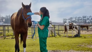
Your vet will start with a static examination of the limb, palpating each sesamoid bone for heat, swelling and pain. The horse is then trotted up in hand on a firm, level surface, and lower limb flexion tests are performed for both forelimbs.
Diagnostic analgesia (nerve blocks)
If lameness is mild and the source is unclear, the vet will block the pain pathways one region at a time. An abaxial sesamoid block usually improves the lameness when the sesamoids or branches of the suspensory are involved.
Radiographs (X-rays)
Three to four standard views of the fetlock are taken, with particular attention to the dorsoproximal-palmarodistal oblique (DP) and lateromedial views. Vets look for enlarged vascular channels, lucent lines, irregular outlines or new bone formation. The pattern and severity allow the case to be graded against the BEVA scheme below.
Ultrasound
Ultrasound assesses the suspensory ligament branches that attach to the sesamoid. Because sesamoiditis often coexists with desmitis of these branches, a combined imaging approach is standard.
Advanced imaging
MRI or nuclear scintigraphy may be recommended for difficult or chronic cases, particularly to identify subtle bone bruising or to differentiate active disease from old, inactive change.
BEVA Radiographic Grading System
UK and European vets typically use the Plevin and McLellan grading system, often referred to in BEVA continuing professional development. The grades describe both the number and the size of vascular channels visible on the proximal sesamoid bones:
| Grade | Findings | Interpretation |
|---|---|---|
| 0 | No vascular channels visible | Normal |
| 1 | 1–2 narrow vascular channels | Mild — within normal range for many horses |
| 2 | 3 narrow channels OR 1–2 wider channels | Mild to moderate — clinical correlation needed |
| 3 | 4+ vascular channels OR 3+ wide channels | Moderate to severe — increased risk of suspensory injury |
| 4 | Confluent or large lytic areas, irregular outline | Severe — guarded prognosis for athletic use |
Grade 3 and 4 findings are increasingly being linked in research papers to a higher chance of subsequent suspensory branch injury, particularly in racehorses, which is why purchasers often baulk at higher-grade radiographs even in the absence of current lameness.
Treatment Options for Sesamoiditis
Treatment is driven by the severity of the bony change, whether the suspensory branches are involved, and the horse’s intended job. The core principles are to control inflammation, support healing and reload the limb gradually.
Rest and controlled exercise
The single most important intervention is rest, but in the modern sense — controlled, gradually loaded movement rather than complete box rest. Horses are typically removed from fast work for 8–24 weeks depending on grade, with hand-walking and small-paddock turnout reintroduced first.
Anti-inflammatory medication
Phenylbutazone (“bute”) is the most commonly prescribed NSAID in the UK and is supplied as a Prescription Only Medicine — Veterinarian (POM-V). It is given at the lowest effective dose for the shortest period needed. Meloxicam (Metacam) is increasingly used for its better gastric tolerance. NSAIDs do not heal the bone — they manage pain so the horse can rest comfortably.
Regenerative medicine
For cases with concurrent suspensory branch desmitis, your vet may suggest:
- Platelet-rich plasma (PRP): Injected into the suspensory branch under ultrasound guidance.
- IRAP (interleukin-1 receptor antagonist protein): Used to dampen inflammation in the surrounding soft tissue.
- Stem cell therapy: Mesenchymal stem cells from the horse’s bone marrow or fat, sometimes used in chronic suspensory branch disease.
- Bisphosphonates (tiludronic acid / clodronic acid): In the UK, Osphos (clodronic acid) is licensed for forelimb lameness associated with bone resorption in the navicular bone, not in horses under 4, and Tildren (tiludronic acid) for bone spavin lameness in horses over 3, not for navicular disease (VMD datasheets). Neither licence covers sesamoid disease, so both are used off-licence in some sesamoid cases under veterinary judgement.
Shockwave therapy
Extracorporeal shockwave therapy is widely used for suspensory branch and sesamoid pain. Treatments are usually given in a series of three sessions, two to three weeks apart.
Physiotherapy and rehab
Veterinary physiotherapy and controlled water-treadmill work, where available, can help maintain proprioception and core strength during the rest period.
Farriery and Shoeing Considerations
Skilled farriery is fundamental to recovery. The aim is to support the suspensory apparatus, ease breakover and rebalance the foot:
- Heel support: Fitting the shoe wide and full at the heels, often with extended branches, helps prevent the heels from collapsing under load.
- Reduced toe length: Shortening the toe and rolling or rocker-shoeing it eases breakover and reduces leverage on the fetlock.
- Mediolateral balance: Regular trim cycles every 5–6 weeks keep the foot from drifting out of balance.
- Pads and packing: Soft pads or pour-in pad material can absorb concussion on hard ground.
- Egg bar or onion shoes: Sometimes used short-term in severely sore horses to maximise support behind the fetlock.
Working with a Registered Farrier (Farriers Registration Council, UK) and ideally a vet-farrier collaboration produces the best outcomes. For horses transitioning to barefoot during rehab, see our guide to barefoot trimming for horses.
Typical Rehabilitation Timeline
Every case is different, but the table below shows the rehab arc many UK vets follow for a moderate (Grade 2–3) case with mild suspensory branch involvement.
| Phase | Duration | Activity | Goal |
|---|---|---|---|
| Acute | Weeks 0–2 | Box rest with daily hand-walking 5–10 min | Reduce inflammation, control pain |
| Early reload | Weeks 3–6 | Hand-walk 15–30 min/day, small-paddock turnout | Maintain limb perfusion, gentle reloading |
| Increased turnout | Weeks 7–12 | Larger paddock, walk under saddle from week 10–12 | Build soft-tissue tolerance to load |
| Re-introduction of trot | Weeks 13–18 | Short trot intervals on a straight line, surfaced school | Test the bone-soft tissue interface |
| Canter and schooling | Weeks 19–24 | Gradual canter work, no fast work or jumping | Build cardiovascular and muscular fitness |
| Return to work | Week 24+ | Discipline-specific work, jumping introduced last | Full return to athletic function if pain-free |
A repeat ultrasound or radiographic check at the 12-week mark is sensible before stepping up to ridden work.
Prognosis and Return to Work
Prognosis depends on grade, age, suspensory involvement and the horse’s intended use:
- Grade 1–2 with no soft-tissue involvement: Excellent — most return to full work without restriction.
- Grade 2–3 with mild suspensory branch desmitis: Good — 60–80% return to previous level of work with patient rehab.
- Grade 3–4 with significant suspensory injury: Guarded — many horses can return to lighter work, but elite performance careers are at risk.
- Bilateral severe sesamoiditis with sesamoid fracture: Poor for racing or top-level jumping; many horses are retired or stepped down to hacking and breeding.
Prevention Strategies for Sesamoiditis
You cannot remove all risk, but you can reduce it significantly:
- Avoid intense work in young, skeletally immature horses. Allow time for the bones to develop before pushing canter work.
- Increase workload gradually — the 10% rule (no more than a 10% increase per week in distance or intensity) suits most disciplines.
- Keep the foot balanced with regular trim cycles every 5–6 weeks.
- Vary the surfaces you work on. Constant deep schooling surfaces can be as damaging as constant hard ground.
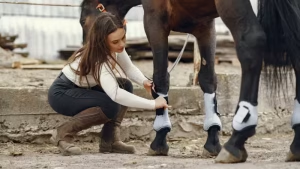
- Use boots or wraps appropriate to the discipline, but be aware they offer concussion management more than mechanical support.
- Maintain a healthy bodyweight. See our guide to horse feeding 101 for managing condition.
- Build core and topline strength with a sensible equine fitness plan.
- Cool down properly after fast work, then check legs for any heat or filling that evening.
Sesamoiditis on Pre-Purchase X-Rays
Many UK pre-purchase examinations now routinely radiograph the fetlocks, and sesamoiditis is one of the most commonly debated incidental findings. A few points worth understanding:
- Grade 1 changes are common in performance horses and not necessarily a deal-breaker.
- Grade 2–3 changes warrant a frank conversation with the vendor and your vet about the horse’s job.
- Bilateral changes are more concerning than unilateral changes, particularly in young Thoroughbreds.
- Always pair the radiographs with a thorough lameness exam and palpation. Pretty pictures with a sound horse are more reassuring than clean pictures with subtle clinical signs.
- Insurance underwriters in the UK often exclude the affected limbs from suspensory and sesamoid cover for the first policy year.
Frequently Asked Questions
How long does sesamoiditis take to heal in horses?
Most mild to moderate cases need 12 to 24 weeks of structured rest and rehab before returning to ridden work. Severe cases or those with significant suspensory ligament involvement may require six to nine months. Repeat imaging at 12 weeks helps guide whether the horse is ready to step up to trotting work.
Can a horse with sesamoiditis be ridden?
Once your vet gives the go-ahead — usually after the acute inflammation has settled and follow-up imaging looks stable — the horse can be ridden under a controlled rehab plan. Walking under saddle is normally introduced first, then trot, then canter. Avoid fast work, jumping or deep ground until your vet confirms the soft tissues have recovered.
What does sesamoiditis look like on an X-ray?
Vets look for enlarged vascular channels (lines running through the bone), lucent areas (darker patches indicating bone loss), an irregular outline of the bone, or fracture lines. The number, width and confluence of these channels determine the BEVA-style grade and guide prognosis.
Is sesamoiditis the same as a sesamoid fracture?
No. Sesamoiditis is inflammation and bony remodelling within the sesamoid, while a sesamoid fracture is a discrete break in the bone. The two can overlap — chronic sesamoiditis can predispose to fracture under fast work — but they are different diagnoses with different prognoses.
How much does it cost to treat sesamoiditis in the UK?
Costs vary widely with severity. A standard work-up (clinical exam, nerve blocks, four radiographs and ultrasound) typically runs from £350 to £700 at most UK first-opinion practices. Add £300–£600 per session for shockwave therapy or PRP, and £600–£1,200 for a course of stem cell therapy. Insurance with vet-fee cover often picks up most of the cost subject to policy excesses.
Can I prevent sesamoiditis returning after rehab?
Yes, in most cases. Stick to a planned, gradual fitness build, maintain regular farriery cycles, vary your training surfaces and avoid sudden spikes in fast work or jumping height. Routine lameness exams and an annual flexion check with your vet help catch low-grade recurrence before it becomes clinical.
Should I buy a horse with Grade 2 sesamoiditis on x-ray?
It depends on the horse’s intended job and current soundness. Many sport horses and leisure horses live and compete happily with Grade 1–2 changes, particularly when the suspensory branches are clean on ultrasound. Discuss the findings with your own vet, factor in the discipline you intend to pursue, and consider how UK insurance underwriters may treat the limb.